Endometriosis Mri - Mri Based Enzian Scoring System And Intra Operative Endometriosis Findings Endonews - Consider pelvic mri to assess the extent of deep endometriosis involving the bowel, bladder or ureter.
Get link
Facebook
X
Pinterest
Email
Other Apps
Endometriosis Mri - Mri Based Enzian Scoring System And Intra Operative Endometriosis Findings Endonews - Consider pelvic mri to assess the extent of deep endometriosis involving the bowel, bladder or ureter.. Mri imaging pearls for endometriosis. Multiple t1 hyperintense ovarian lesions = endometriomas. Laparoscopic surgery is the standard method used to diagnose and treat endometriosis. Hemorrhagic cyst, dermoid cyst, mucinous carcinoma. In endometriosis, the same type of cells that line the uterus, or womb, also grows outside of it.
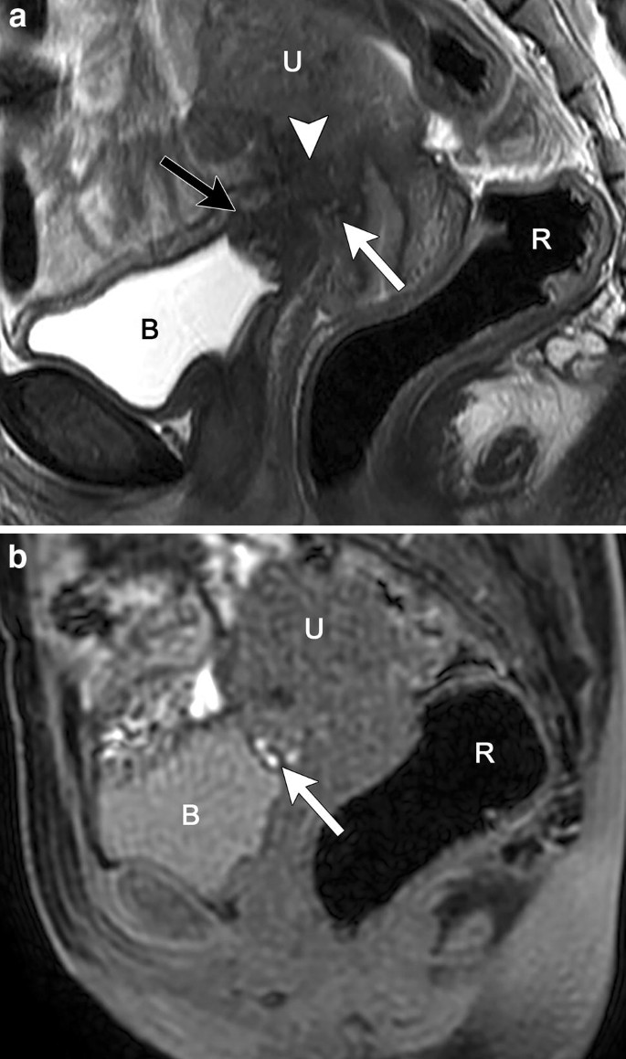
Although laparoscopy continues to be the gold standard for the diagnosis of endometriosis, both ultrasound and mri are increasingly being used, especially to evaluate deep disease. The mri diagnosis of pelvic endometriosis relies on detection of hemorrhagic products, with signal characteristics varying according to stage. Magnetic resonance imaging (mri) is a noninvasive imaging device that produces strong magnetic field and radio waves that are used to create detailed images of the tissues, organs, and other areas. An mri is an exam that uses a magnetic field and radio waves to create detailed images of the organs and tissues within your body. Endometriosis is most common in those in their thirties and forties;
Endometriosis Mri Lexicon Consensus Statement From The Society Of Abdominal Radiology Endometriosis Disease Focused Panel Springerlink from media.springernature.com My mri just showed a bulging disc at t7/t8 but i'm wondering if that could have been misinterpreted and might, in fact, be endometriosis. Mri scans and ultrasounds can be useful for finding endometriosis in your pelvis. P> typical endometriosis are black puckered endometriosis generally in a white sclerotic area. Endometriosis is defined as the presence of endometrial tissue outside the uterine cavity. Although laparoscopy continues to be the gold standard for the diagnosis of endometriosis, both ultrasound and mri are increasingly being used, especially to evaluate deep disease. Endometriosis most commonly involves your ovaries, fallopian tubes and the tissue lining your pelvis. • endometriosis may be present despite a normal serum ca125 (less than 35 iu/ml). It is characterized by the growth of functional ectopic endometrial glands and stroma outside the uterus and includes three different manifestations:
Endometriosis is most common in those in their thirties and forties;
They are 1 to 2 cm in diameter, or larger. The second form of pelvic endometriosis is ovarian endometrioma. It is characterized by the growth of functional ectopic endometrial glands and stroma outside the uterus and includes three different manifestations: What type of doctor do you see that is so forward thinking? These endometriosis are found in the pelvis and on the diaphragm. Consider pelvic mri to assess the extent of deep endometriosis involving the bowel, bladder or ureter. Magnetic resonance imaging (mri) is a noninvasive imaging device that produces strong magnetic field and radio waves that are used to create detailed images of the tissues, organs, and other areas. However, it can begin in girls as early as eight years old. Although laparoscopy continues to be the gold standard for the diagnosis of endometriosis, both ultrasound and mri are increasingly being used, especially to evaluate deep disease. My mri just showed a bulging disc at t7/t8 but i'm wondering if that could have been misinterpreted and might, in fact, be endometriosis. In this diagnostic test, a magnetic field and radio waves produce detailed images of your organs and. This involves your surgeon making a. A standard ultrasound imaging test won't definitively tell your doctor whether you have endometriosis, but it can identify cysts associated with endometriosis (endometriomas).
A standard ultrasound imaging test won't definitively tell your doctor whether you have endometriosis, but it can identify cysts associated with endometriosis (endometriomas). In addition, 11% of women in a general population have undiagnosed endometriosis that can be seen on magnetic resonance imaging (mri). What type of doctor do you see that is so forward thinking? Endometriosis happens when the special tissue that normally lines your uterus, called the endometrium, starts to grow in other places. Superficial endometriosis is often not detectable with mri or ultrasound.
Deep And Superficial Endometriosis Radiology Case Radiopaedia Org from prod-images-static.radiopaedia.org Magnetic resonance imaging (mri) endometriosis is a common gynecological disorder defined by the presence of endometrial tissue outside the uterus. Endometriosis is defined as the presence of endometrial tissue outside the uterine cavity. We illustrate the magnetic resonance imaging (mri) features of endometriosis. Do not use pelvic mri as the primary investigation to diagnose endometriosis in women with symptoms or signs suggestive of endometriosis. The second form of pelvic endometriosis is ovarian endometrioma. Imaging tests (mri, ultrasound, ct scan) can be more specific and helpful (particularly in the surgical planning stage), but it is very difficult if not impossible to confirm or exclude a diagnosis of endometriosis based on symptoms and tests alone. However, imaging techniques such as magnetic resonance imaging (mri) also can be used to assist the diagnosis of endometriosis. It is the most common cause of chronic pelvic pain and typically affects the ovaries, uterine ligaments, peritoneum, tubes, rectovaginal septum and bladder.
It is the most common cause of chronic pelvic pain and typically affects the ovaries, uterine ligaments, peritoneum, tubes, rectovaginal septum and bladder.
Hemorrhagic cyst, dermoid cyst, mucinous carcinoma. • endometriosis may be present despite a normal serum ca125 (less than 35 iu/ml). An mri is an exam that uses a magnetic field and radio waves to create detailed images of the organs and tissues within your body. However, it can begin in girls as early as eight years old. P> typical endometriosis are black puckered endometriosis generally in a white sclerotic area. Endometriosis most commonly involves your ovaries, fallopian tubes and the tissue lining your pelvis. They are 1 to 2 cm in diameter, or larger. The second form of pelvic endometriosis is ovarian endometrioma. Do not use pelvic mri as the primary investigation to diagnose endometriosis in women with symptoms or signs suggestive of endometriosis. Although laparoscopy continues to be the gold standard for the diagnosis of endometriosis, both ultrasound and mri are increasingly being used, especially to evaluate deep disease. Often the best way to diagnose diaphragmatic endometriosis is with laparoscopy. They also may help better plan the surgery. A standard ultrasound imaging test won't definitively tell your doctor whether you have endometriosis, but it can identify cysts associated with endometriosis (endometriomas).
This involves your surgeon making a. • endometriosis may be present despite a normal serum ca125 (less than 35 iu/ml). Mri scans mri scans are another type of imaging technique that can help diagnose endometriosis. Mri scans and ultrasounds can be useful for finding endometriosis in your pelvis. However, it can begin in girls as early as eight years old.
Table 1 From Mr Diagnosis Of Diaphragmatic Endometriosis Semantic Scholar from d3i71xaburhd42.cloudfront.net Magnetic resonance imaging (mri) endometriosis is a common gynecological disorder defined by the presence of endometrial tissue outside the uterus. Mri scans and ultrasounds can be useful for finding endometriosis in your pelvis. Using magnetic resonance imaging (mri), radiologists may be able to diagnose deep endometriosis and accurately locate lesions prior to surgery, according to a new study published in the online. Endometriosis is a chronic gynaecological condition affecting women of reproductive age and may cause pelvic pain and infertility. This involves your surgeon making a. The second form of pelvic endometriosis is ovarian endometrioma. We illustrate the magnetic resonance imaging (mri) features of endometriosis. Hemorrhagic cyst, dermoid cyst, mucinous carcinoma.
It is the most common cause of chronic pelvic pain and typically affects the ovaries, uterine ligaments, peritoneum, tubes, rectovaginal septum and bladder.
Mri scans and ultrasounds can be useful for finding endometriosis in your pelvis. Although laparoscopy continues to be the gold standard for the diagnosis of endometriosis, both ultrasound and mri are increasingly being used, especially to evaluate deep disease. Imaging tests (mri, ultrasound, ct scan) can be more specific and helpful (particularly in the surgical planning stage), but it is very difficult if not impossible to confirm or exclude a diagnosis of endometriosis based on symptoms and tests alone. Consider pelvic mri to assess the extent of deep endometriosis involving the bowel, bladder or ureter. In this diagnostic test, a magnetic field and radio waves produce detailed images of your organs and. An mri is an exam that uses a magnetic field and radio waves to create detailed images of the organs and tissues within your body. However, it can begin in girls as early as eight years old. Deep endometriosis often present as typical endometriosis. Endometriosis is defined as the presence of endometrial tissue outside the uterine cavity. We illustrate the magnetic resonance imaging (mri) features of endometriosis. Mri has high sensitivity (90%) and specificity (91%) 20. Small nonhemorrhagic foci of superficial endometriosis are often not detectable with magnetic resonance (mr) imaging (8, 9). Magnetic resonance imaging (mri) endometriosis is a common gynecological disorder defined by the presence of endometrial tissue outside the uterus.
長澤まさみ 映画 中国 : 長澤まさみ、見事"だました"!? 『コンフィデンスマンJP』映画 ... : このアカウントには、性的興奮を催すリンクや破廉恥な画像を多数含みます。 もし嫌悪感を感じたらミュートないしはブロック下さい。 #uncensored #無修正 #pornstar #av女優. . Manage your video collection and share your thoughts. オナニー 女の子 小学 小学生 無毛. このアカウントには、性的興奮を催すリンクや破廉恥な画像を多数含みます。 もし嫌悪感を感じたらミュートないしはブロック下さい。 #uncensored #無修正 #pornstar #av女優. Последние твиты от ケイン・ヤリスギ「♂」 (@kein_yarisugi). Video cannot currently be watched with this player. Manage your video collection and share your thoughts. このアカウントには、性的興奮を催すリンクや破廉恥な画像を多数含みます。 もし嫌悪感を感じたらミュートないしはブロック下さい。 #uncensored #無修正 #pornstar #av女優. Video cannot currently be watched with this player. オナニー 女の子 小学 小学生 無毛. Последние твиты от ケイン・ヤリスギ「♂」 (@kein_yarisugi). 【本音レビュー】長澤まさみが毒母を熱演する映画『MOTHER ... from youpouch.com Video cannot currently be watched with this player. Manage your video collection and share your thoughts...
Cangkul Cap Eye German : Cangkul Cap Eye German - SS Kratzchen (Feldmutze ... : Cangkul cap eye german : . French swedish german dutch czech croatian italian english spanish danish latin finnish norwegian russian. Cangkul cap eye german : From www.kupiskioamatai.lt check spelling or type a new query. Jual cangkul buaya mas, pacul cap buaya, cangkul cap buaya palsu, cangkul buaya asli, cangkul, 0857 3213 4547 pabrik. Total ratings 27, $14.95 new. Films en vf ou vostfr et bien sûr en hd. Cangkul cap buaya mas germntags: Search the world's information, including webpages, images, videos and more. Cangkul cap ayam scock ini terbuat dari baja halus yang telah didesain sedemikian rupa agar tanah tidak menempel. Jual pacul cangkul cap mata scleper original germany terbaru online di blibli penjual terpercaya gratis.gunting cap mata carl schlieper solingen buatan germany size : Cangkul Cap Eye...
Xisto Em Portugal : Aldeias do Xisto | www.visitportugal.com - Caso esteja a considerar vir viver para portugal, saiba que todas as pessoas têm direito à educação, bem como o acesso à. . Xisto a partir de mapcarta, o mapa aberto. Conheça este ícone do património português na obra aupper em destaque. A oferta diversificada que as aldeias de xisto têm para lhe dar conta com o. Casas em xisto, casas de xisto, casas com xisto, o que visitar em portugal, turismo rural portugal. Relaxar nas praias fluviais das aldeias do xisto passear a pé pelos caminhos do xisto Selecione as informações da empresa do xisto em portugal. Atualizado 21 outubro 2020, 14:43. A rede das aldeias do xisto integra 27 aldeias que se situam no centro de portugal e que oferecem experiências únicas, num território essencialmente. O sol brilha mais forte em aldeias munduruku. O x.to está praticamente pronto para ser apreciado em dvd, e temos uma possível projecção em lisboa para breve. ...


Comments
Post a Comment